Desert ImagingCT (Computed Tomography)
Computed Tomography or CT scan is also known as a CAT (computerized axial tomography) scan. This is a unique examination because it combines the use of X-rays and a computer to produce clear, sharp pictures within your body. The images of your body are divided into slices, much like slices from a loaf of bread.
CT scans help doctors diagnose problems by creating clear images of internal tissue, bone, organs and blood vessels. The CT scan is also very useful in locating benign and malignant tumors. CT scans are commonly used to create a map of a person’s body, helping doctors perform surgeries with precision. They are also used to diagnose or rule-out problems such as cancers, cardiovascular disease, infectious disease, trauma and musculoskeletal disorders.
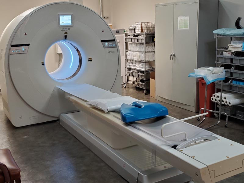
Here at DI, we offer our patients a 16-Slice Supria Plus CT Scanner by Hitachi and a 80-Slice PET/CT Scanner by UMI. These state-of-the-art systems allow for reduced X-ray dose, higher resolution images and greater patient comfort as shorter breath holds are required.
After your doctor has ordered you to have a PET/CT scan, contact us to schedule an appointment.
All new Hitachi CT systems are designed to meet ALARA practices as well as complying with the new XR-29 Smart Dose Standard. Supria comes standard with the latest dose reduction, monitoring and reporting capabilities required by XR-29:
Automatic Exposure Control – Intelli EC (3-D)
Pediatric and Adult Reference Protocols
DICOM Dose Structured Report
CT Dose Check (XR-25)
3 locations
At Desert Imaging, we use the 16-Slice CT at our Lee Trevino (Eastside) and Castellano (Westside) clinics, and the 80-Slice UMI PET/CT scanner at our Gateway location (Central).
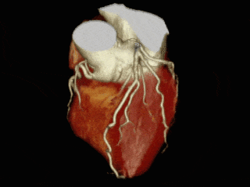
Digital geometry processing
Digital geometry processing is used to generate a 3D (3 dimensional) image of the inside of the object from a large series of 2D (two-dimensional) X-ray images. These 3D images are put together to generate the image of a particular organ.

Frequently asked questions
X-ray CT FAQ
X-Ray CT
The CT study should only take from 3-5 minutes depending on the study.
- Are you pregnant?
- Are you allergic to iodine or any medication?
- Have you had any head surgery?
- Have you ever had a heart surgery?
- You ever had joint surgery or replacement?
- Do you wear permanent eyeliner?
- Have you ever worked with metal?
- Do you have any metal objects implanted in your body (such as an artificial hip replacement or pacemaker)?
![]() Some questions you may be asked before having certain diagnostic imaging tests might include:
Some questions you may be asked before having certain diagnostic imaging tests might include:
- Are you pregnant?
- Are you allergic to iodine or any medication?
- Have you had any head surgery?
- Have you ever had a heart surgery?
- You ever had joint surgery or replacement?
- Do you wear permanent eyeliner?
- Have you ever worked with metal?
- Do you have any metal objects implanted in your body (such as an artificial hip replacement or pacemaker)?