Desert ImagingPET/CT SCAN
What is aPET/CT Scan?
PET/CT is a powerful imaging tool that combines a PET (positron emission tomography) scan and a computed tomography (CT) scan. PET/CT is used to diagnose, stage, or restage cancer, as well as evaluate the effectiveness of treatment. The images provide information about the location, nature, and size of a tumor or mass. The CT’s newer technology and faster scan times result in less radiation exposure to patients. Its wider open design decreases claustrophobia and provides better access for larger patients and radiation therapy planning for cases requiring more space, such as breast cancer.
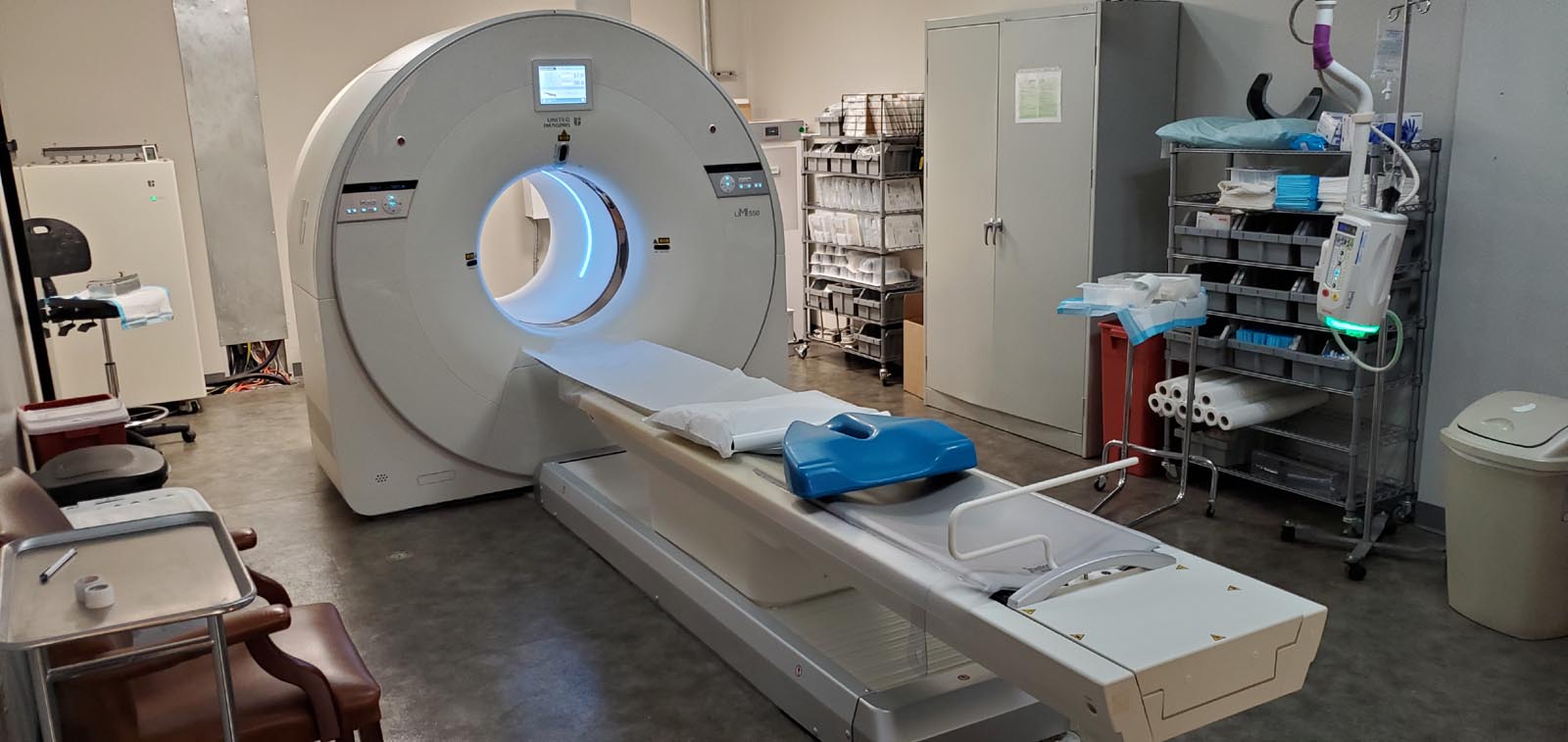
At Desert Imaging, we use a 80-slice PET/CT scanner by UMI, which features the most advanced technology in the greater El Paso region. During the exam, the patient is first injected with a glucose (sugar) solution that contains a tracer. The tracer is absorbed by the particular organs or tissues being examined. The PET/CT scanner is then able to "see" damaged or cancerous cells where the glucose is absorbed (cancer cells use more glucose than healthy cells). The exam is painless and takes approximately 30 minutes to complete.
Desert ImagingPET/CT Exams
Brain
Cardiac (13N Ammonia)
DOTATATE (Ga-68)
Limited
Prostate Specific Membrane Antigen (PSMA)
Whole Body
Scheduling your exam
After your doctor has ordered you to have a PET/CT scan, contact us to schedule an appointment.
A PET/CTstudy helps your physician diagnose problems, pinpoint the best approach to treatment and monitor your progress.
BecauseThe sooner you know,
The better

Desert Imaging is the only center in the region offering a 80 slice PET/CT scan
PET + CT
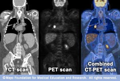
Combining a PET scan with an MRI or CT scan help make the images easier to interpret. The image on the far left is a CT scan, while the center image is from a PET scanner. The image on the right is a combined PET/CT scan. This image clearly shows where the bright spot on the chest is located and where that particular incidence of lung cancer is located in relation to other organs.
The wealth of information that a scan provides may enable your doctors to confirm that your treatment is working, or switch you to a more effective treatment immediately.
What is Cardiac PET MPI?
PET – Positron Emission Tomography
MPI – Myocardial Perfusion Imaging
A Cardiac PET/CT MPI scan provides accuracy, excellent quality, and the ability to detect disease before symptoms are present. This exam can diagnose cardiac diseases with greater certainty and lower radiation exposure than traditional stress testing and is the gold standard for diagnosing coronary heart disease.We use this exam to evaluate the health of your heart by measuring the blood flow it receives. The results of this exam determine if treatment is needed or to manage your current treatment.
What to EXPECT?
Most cardiac PET exams take less than one hour; however, a longer exam should not be cause for concern. Upon arrival, our team will register you and review your medical history. Please be sure to report all medications you are taking.Trained medical personnel will be with you throughout your exam.An intravenous line will be placed in a vein in your arm to allow the administration of medication during the exam.Small pads called electrodes will be placed on your chest to monitor the electrical activity of your heart through the study. Your blood pressure, heart rate, and electrocardiogram are monitored before, during, and after the test.You will be asked to lie down on a padded, comfortable scanning table developed especially for the PET camera. During your PET scan, most of your body is outside the scanner; this is not a full-body scan. If you’re claustrophobic, please take your medication 30 minutes before your scan.A small amount of radiopharmaceutical is given through your intravenous line, enabling the PET camera to capture images of your heart. The camera will take pictures of your heart in two phases: one at rest and one at stress.The resting and stress phases are compared to evaluate blood flow through your heart and to look for heart disease and damage to the heart muscle.
Why Choose Cardiac PET MPI?
Patients benefit from a Cardiac PET MPI exam for a variety of reasons:
Breast implants, large chest
High-risk patients
Known or suspected coronary artery disease
Large body habitus, high BMI
Prior poor-quality stress imaging study
Requires pharmacologic stress imaging
Time-sensitive, urgent
Unable to exercise
Young patients with coronary artery disease
The need for a more accurate test
Safety
Cardiac PET MPI scans are a safe and effective diagnostic imaging test. The tracer used is not a dye or contrast and results in minimal radiation exposure and enables a far briefer test.
Frequently asked questions
PET/CT SCAN FAQ
PET/CT SCAN
A positron emission tomography (PET) scan is an imaging test that uses a radioactive substance called a tracer to look for disease in the body. A PET scan shows how organs and tissues are working.
![]()
- Make sure you haven't had anything to eat 6 hours prior to your appointment.
- No cough drops, cough syrup, breath mints, chewing gum, or even "sugar-free" stuff.
- Check with your doctor if you are diabetic. Blood sugar needs to be below a certain level during the exam to avoid any misreading.