Desert ImagingCardiovascular
What is Cardiac PET MPI?
PET – Positron Emission Tomography
MPI – Myocardial Perfusion Imaging
A Cardiac PET/CT MPI scan provides accuracy, excellent quality, and the ability to detect disease before symptoms are present. This exam can diagnose cardiac diseases with greater certainty and lower radiation exposure than traditional stress testing and is the gold standard for diagnosing coronary heart disease.We use this exam to evaluate the health of your heart by measuring the blood flow it receives. The results of this exam determine if treatment is needed or to manage your current treatment.
What to EXPECT?
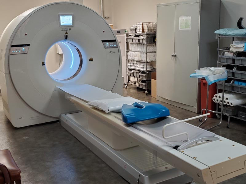
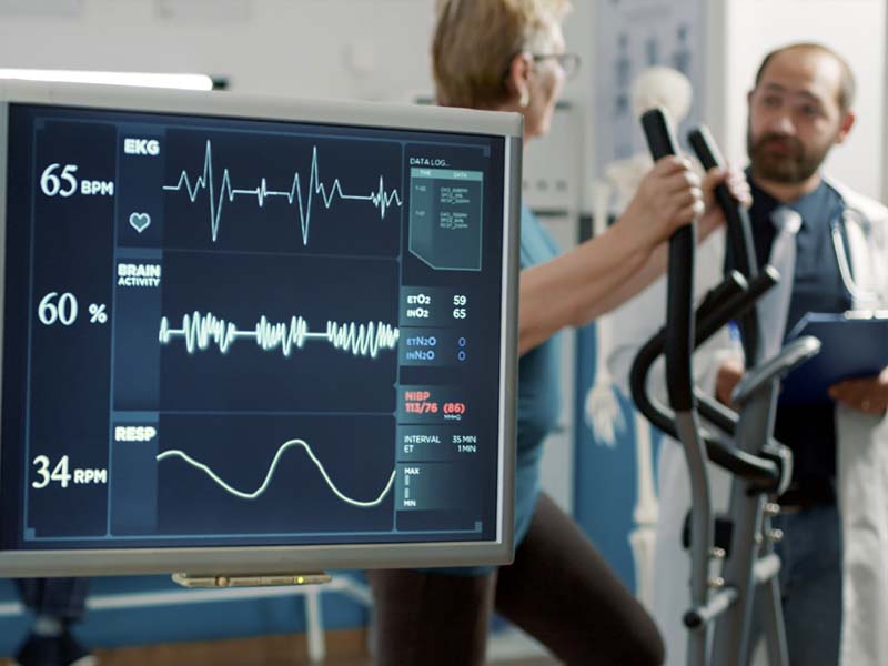
Most cardiac PET exams take less than one hour; however, a longer exam should not be cause for concern. Upon arrival, our team will register you and review your medical history. Please be sure to report all medications you are taking.Trained medical personnel will be with you throughout your exam.An intravenous line will be placed in a vein in your arm to allow the administration of medication during the exam.Small pads called electrodes will be placed on your chest to monitor the electrical activity of your heart through the study. Your blood pressure, heart rate, and electrocardiogram are monitored before, during, and after the test.You will be asked to lie down on a padded, comfortable scanning table developed especially for the PET camera. During your PET scan, most of your body is outside the scanner; this is not a full-body scan. If you’re claustrophobic, please take your medication 30 minutes before your scan.A small amount of radiopharmaceutical is given through your intravenous line, enabling the PET camera to capture images of your heart. The camera will take pictures of your heart in two phases: one at rest and one at stress.The resting and stress phases are compared to evaluate blood flow through your heart and to look for heart disease and damage to the heart muscle.
Why Choose Cardiac PET MPI?
Patients benefit from a Cardiac PET MPI exam for a variety of reasons:
Breast implants, large chest
High-risk patients
Known or suspected coronary artery disease
Large body habitus, high BMI
Prior poor-quality stress imaging study
Requires pharmacologic stress imaging
Time-sensitive, urgent
Unable to exercise
Young patients with coronary artery disease
The need for a more accurate test
Safety
Cardiac PET MPI scans are a safe and effective diagnostic imaging test. The tracer used is not a dye or contrast and results in minimal radiation exposure and enables a far briefer test.
STRESS TEST
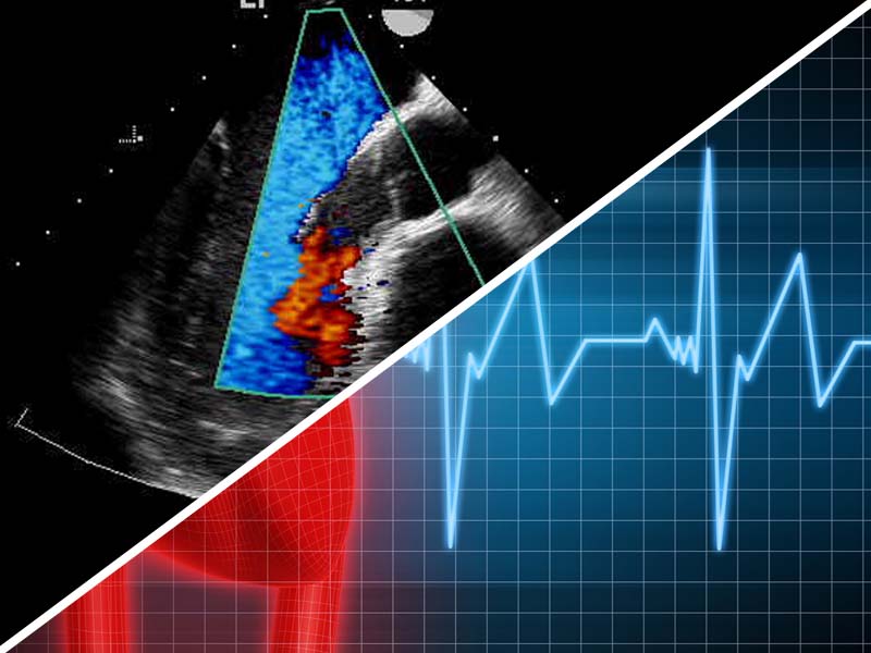
A stress test helps determine if chest pain or other symptoms a patient is feeling are related to heart disease. It utilizes an echocardiogram, also known as a stress echo or SE. This ultrasound imaging of the heart is used to assess the heart wall motion in response to physical stress. First, images of the heart are taken "at rest" to acquire a baseline of the patient's heart wall motion at a resting heart rate. The patient then walks on a treadmill or utilizes another exercise modality to increase the heart rate to his or her target heart rate. Finally, images of the heart are taken "at stress" to assess heart wall motion at the peak heart rate. A stress echo assesses wall motion of the heart; it does not, however, image the coronary arteries directly.
Echocardiogram
An echocardiogram is an ultrasound of the heart and is referred to as an echo. It can utilize 2D, 3D, and Doppler to create images of the heart. It is used to diagnose, manage, and follow up patients suspected or having problems with their heart.
EKG
An EKG is an electrocardiogram. It looks at the electrical activity of the heart using electrodes placed on the patient’s body.
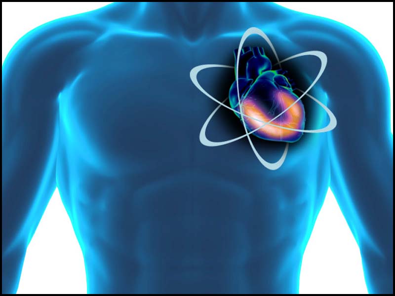
Nuclear Cardiology
During a nuclear cardiology test, a very small amount of radioactive tracer (radionuclide) is injected into a vein and is taken up by the heart. A very sensitive gamma camera then takes still pictures and movies of the heart with rest, exercise, or medication-induced stress testing.
Calcium Scoring
Coronary calcium scoring is also called cardiac calcium scan. The coronary arteries supply blood to the heart. Normally, the coronary arteries do not contain calcium. Calcium in the coronary arteries may be a sign of coronary artery disease. This study is done with the 80 slice CT scan.
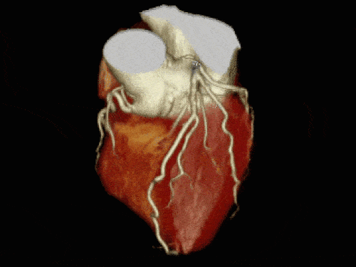
80 Slice CT of the Heart
The 80-slice CT scan provides dramatic detailed images of your coronary arteries and heart in just minutes, and without requiring an invasive and expensive hospital-based cardiac catheterization. Ground-breaking advances in heart imaging technology are now within reach, making possible early and rapid diagnosis of heart disease.