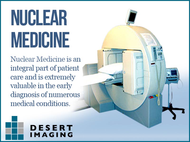
Desert ImagingNuclear Medicine
How it works
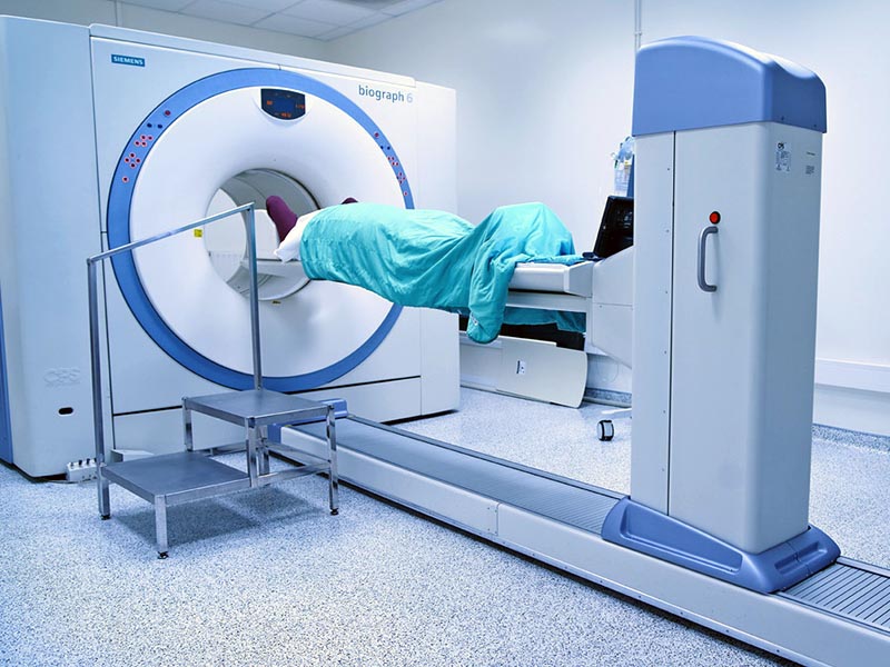
Nuclear medicine is a branch of radiology that involves administering small amounts of radioactive material (called radiopharmaceuticals or radiotracers) to a patient by injection, inhalation, or pill. The radiopharmaceutical eventually accumulates in a particular organ or area of the body, where it gives off energy in the form of gamma rays. The energy is detected by a special camera, which produces a series of images on a computer screen or film. This provides us with details about the structure and function of an organ, tissue or bone in the body. This test also allows us to identify abnormalities early in the progression of a disease.
Nuclear medicine imaging
We use nuclear medicine imaging to diagnose and treat diseases that affect the heart, brain, kidney, bladder, thyroid, liver, lung and bone cancers.
Some types of nuclear medicine tests we offer include:
- Bone scan
- Hepatobiliary (HIDA) Scan
- Lung Scan
- Multiple-Gated Acquisition (MUGA) scan
- Renal Scan
- Thyroid Uptake and Scan
- Thyroid Treatment
- Cardiac Perfusion Scan
- Gastric Emptying Study
- Liver/Spleen Scan
- G.I. Bleed Study
Nuclear medicine is a medical specialty that is used to diagnose and treat diseases in a safe and painless way. Nuclear medicine procedures permit the determination of medical information that may otherwise be unavailable, require surgery, or necessitate more expensive and invasive diagnostic tests. The procedures often identify abnormalities very early in the progression of a disease — long before some medical problems are apparent with other diagnostic tests. This early detection allows a disease to be treated sooner in its course when a more successful prognosis may be possible.
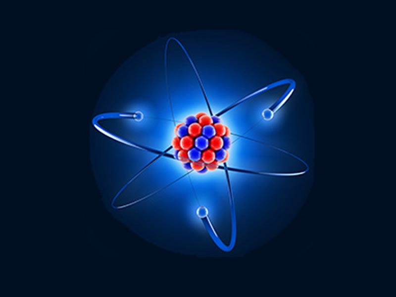
Nuclear medicine refers to medicine (a pharmaceutical) that is attached to a small quantity of radioactive material (a radioisotope). This combination is called a radiopharmaceutical. There are many different radiopharmaceuticals available to study different parts of the body. Which radiopharmaceutical is used will depend upon the condition to be diagnosed or treated.

Why is it called nuclear medicine?
Radiopharmaceuticals are introduced into the patient’s body by injection, swallowing, or inhalation. The amount given is very small. The pharmaceutical part of the radiopharmaceutical is designed to go to a specific place in the body where there could be disease or an abnormality. The radioactive part of the radiopharmaceutical that emits radiation, known as gamma rays (similar to x-rays), is then detected using a special camera called a gamma camera. This type of camera allows the nuclear medicine physician to see what is happening inside your body. During this imaging procedure, the patient is asked to lie down on a bed and then the gamma camera is placed a few inches over the patient’s body. Pictures are taken over the next few minutes. These images allow expert nuclear medicine physicians to diagnose the patient’s disease.
How do radiopharmaceuticals work?
Although exposure to radioactivity in very large doses can be harmful, the radioactivity in radiopharmaceuticals is carefully selected by the nuclear medicine physician to be safe. Yes. There are several types of gamma cameras, small and large. Depending upon the kind of pictures that need to be taken, these cameras will operate in a stationary mode, move across the body or rotate around the body. Gamma cameras do not hurt, nor do they make any noise that might frighten patients. Also, unlike other imaging devices, such as CT scanners, ultrasound and MRI, gamma cameras do not transmit any radiation to the patient. Do radiopharmaceuticals hurt when they are given? Not at all. They are given in a very small dose, just a few drops. Absolutely. Like any medicine, they are prepared with great care. Before they are used, they are tested carefully and are approved for use by the U.S. Food and Drug Administration. The quantity of the pharmaceutical part of the radiopharmaceutical is very small, generally 1/10th of a millionth of an ounce. The risk of a reaction is 2-3 incidents per 100,000 injections, over 50% of which are rashes, as compared to 2000-3000 per 100,000 injections of x-ray contrast media.
Frequently asked questions
Did you know?
NUCLEAR MEDICINE IMAGING
We use nuclear medicine imaging to diagnose and treat diseases that affect the heart, brain, kidney, bladder, thyroid, liver, lung and bone cancers.
![]()
- Arrive at least 15 minutes before your scheduled appointment time.
- Bring all health insurance cards and information.
- Notify us if any of your information has changed
- Wear loose, comfortable clothing
- Leave valuable items at home